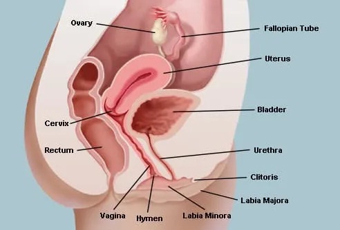
Female Internal Genital Organs
Written by Dr.M.D.Mazumdar, MD
The vagina, the uterus, the two fallopian tubes and the two ovaries constitute the internal genital organs of the female body. They cannot be seen without using special methods. The vagina can be partially seen when the feet are held wide apart and the labia majora and the labia minora are opened up.
In virgins, the vaginal opening is partially closed by a hymen and the vagina cannot be easily seen. In women who have had sexual intercourse, the vagina can be seen by opening the vaginal aperture.
In older women, especially in women after the menopause, the vaginal opening may gape open and the vagina readily examined without any special method.
The Vagina
The vagina is an elastic, fibromuscular, hollow structure extending upwards and backwards from the vulva.
It connects the uterus to the external surface of the body through the vaginal opening in the vulva. The cervix of the uterus protrudes into the vagina at its upper end.
The vagina is the organ for sexual intercourse in the female and forms the birth canal in labour during the delivery of a baby. It is also the opening through which the menstrual blood flows out during the menstrual period.
It is about 2.5 cm wide and 7 cm to 9cm long. But it has a great capacity to distend as is seen during childbirth.
G-Spot
The G-spot or the Graafenberg spot (also spelt as 'Grafenberg') is believed to be an erotic spot in the vagina that is highly sensitive to sexual stimulus. It is said to be a triangular area in the anterior vaginal wall, about 2.5cm from the vaginal opening and along the urethra. But anatomical dissection by many researchers have not found evidence of the existence of the G-spot.
There is increasing evidence which suggests that the G spot lies right over a deep part of the clitoris. Pressure over the G spot stimulate this deeper part of the clitoris and causes greater sexual pleasure.
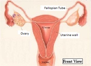
Uterus
The uterus is a hollow, muscular pear-shaped structure situated deep in the pelvis and well protected by the pelvic bones.
When viewed from the front, it looks roughly like an inverted triangle with the broad base as its roof and the narrow apex at its lower end. This lower end also called the cervix opens into the vagina. The two angles on the two ends of the base of the triangle is connected to the two Fallopian tubes.
The adult uterus is about 8cm long and 5cm wide at its thickest part. Before the onset of menstruation, its length ranges from 2cm to 5 cms. The weight varies from 50gms to 80 gms. The non-pregnant uterus can hold only about 5ml of fluid.
The inner lining of the uterus - called the 'endometrium' - is a spongy area with a rich blood supply. Monthly shedding of this endometrium results in the phenomenon called 'menstruation'.
At full term pregnancy, the weight of the uterus can increase to roughly 1 kg and it not only holds the baby but also the placenta and the amniotic fluid surrounding the baby.
The muscles of the uterus have a great capacity to increase both in number and in size (up to 500 times its normal size) when pregnancy occurs. The uterus is the organ that receives, implants, retains and nourishes the fetus till it is mature. It increases gradually in size from the first trimester through the second trimester and upto the end of the third trimester. It then contracts and expels the foetus at the time of labour and childbirth.
Fallopian Tubes
Also called the 'Oviducts', there are two fallopian tubes on either side of the uterus. They extend out from the uterus like arms, reaching for the ovaries which are positioned one on each side of the uterus. Each tube has two openings. One opening connects to the uterus. The other opening is larger and wider and has a number of finger-like projections all around it called the 'fimbriae'.
The fimbriae lie near the ovary of the same side and picks up the ovum at the time it is released from the ovary ('ovulation'). Microscopic hairs called cilia line the inner side of the tube and help in propelling the ovum towards the uterus.
Each tube is about 10cms long. The width varies at different parts along the length, being more towards the ovarian side and thinner but more muscular towards the uterine side. Its widest part, the ampulla lies next to the fimbria and its importance lies in the fact that fertilization of the ovum by the sperm usually occurs in this region.
The fallopian tubes are also the commonest site where blockage can occur leading to infertility.
Ovaries
The two ovaries are situated on either side of the uterus. They are the female sex gonads and are responsible for the release of a mature ovum every month ('ovulation'). They are also the chief producers of the female sex hormones - estrogen and progesterone.
The ovaries are pinkish-white in colour and roughly oval in shape, being 3cm in length, 2cm in breadth and 1cm in thickness approximately. Each ovary has an outer thick lining called the 'cortex' and an inner part called the 'medulla'.
During the reproductive life, i.e. from puberty to menopause, the cortex contains numerous 'Graafian follicles' at different stages of development. Every month a Graafian follicle in one of the ovaries matures and releases an ovum. This phenomenon is called 'ovulation'. During a woman's lifetime, only about 400 follicles reach maturity.
A woman attains menopause when the number of follicles in her ovaries decreases below a critical level. The ovaries shrink in size and become whitish in colour. They also secrete lesser amounts of estrogen and progesterone.
Read More:


Also Read-
- Normal Vaginal Discharge .
- How Pregnancy Occurs.
- Different Sexually Transmitted Diseases .
- Various Methods of Birth Control.
Do you have a gynecological or obstetrical problem? Would you like to discuss it in private? Consult our online gynecologist Dr.M.D.Mazumdar, MD (O&G), at any time you want and get your reply within 24 hours.We charge a nominal fee of USD 20 ($20) per question through Paypal.com.
The procedure of asking a question is quite simple. Clicking on the link below takes you to the Paypal website where the payment is made. After the payment goes through, you will be directed back to this website where you can ask your question. And rest assured, you will get your answer within 24 hours. And usually, even sooner.